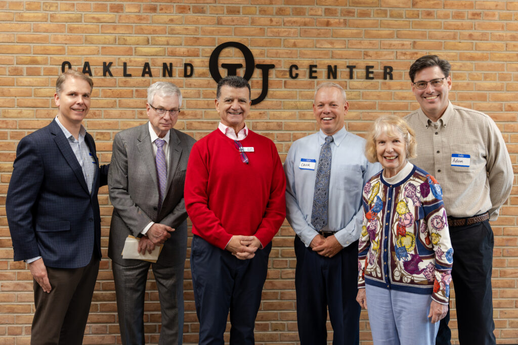
The PRRF Family Connection Conference held both virtually and at Oakland University’s Oakland Center in Rochester, Michigan, brought together PRRF board members, families affected by rare pediatric retinal diseases, and leading pediatric retina specialists.
The opportunity to “Ask a Clinician” all your burning questions is one of the most anticipated sessions during the Pediatric Retinal Research Foundation’s annual Family Connection Conference. Each year, participants bring questions that often have been on everyone’s minds. It sparks a dialogue that helps families stay informed but also reminds us how deeply we are connected.
This year we hosted an extraordinary conversation with PRRF Board President and pediatric retina specialist, Dr. Antonio Capone, Jr., and board member and ophthalmologist Dr. Matthew Trese. They covered everything from Familial Exudative Vitreoretinopathy (FEVR) and Retinopathy of prematurity (ROP) to gene therapy, clinical trials, and the future of personalized medicine for pediatric retinal diseases. Dr. Patrick Droste, pediatric ophthalmologist, also joined the conversation to share his decades of expertise on visual rehabilitation.
Below are the highlights. Watch the full Q&A session on YouTube →
FEVR (Familial Exudative Vitreoretinopathy): Retinal Tears, Treatment & Genetic Testing
The session opened with a question from a participant on Zoom, asking whether retinal tears are a normal finding in young adults with FEVR.
Dr. Capone explained that in the United States, FEVR almost universally causes tractional retinal detachments, driven by scar tissue contracting on the retina, not tears. However, in parts of Asia, a combination of both traction and tears is more common, which has even led researchers in Japan to propose different classification systems for FEVR.
💡 Key Insight: If your child with FEVR in the US has retinal tears, Dr. Capone recommends discussing whether another condition, such as high myopia (nearsightedness), might also be contributing.
Treatment Options for FEVR Retinal Tears and Holes
Dr. Trese walked the audience through how tears and holes are managed, depending on how early they’re caught:
- Prophylactic laser can treat at-risk areas of the retina before a hole or tear develops in patients with familial exudative vitreoretinopathy.
- If a hole is caught before fluid accumulates, laser or cryotherapy (a freezing treatment) can seal the retina in place, preventing a detachment.
- If fluid has already caused a retinal detachment, the fluid must first be drained, the retina repositioned, and then the weld applied.
- In some cases, a scleral buckle (an external support device) may also be needed.
Why Genetic Testing Matters for Every FEVR Family
Both doctors were emphatic: genetic testing is essential for FEVR patients, even if the results don’t always return positive. Knowing the specific gene involved helps predict the clinical course, shapes how closely a patient should be followed, and can reveal whether siblings or parents also carry the gene, even if they have perfectly normal vision.
💡 Family Spotlight: It’s not uncommon for a child to have a severe FEVR presentation while the parent who passed on the gene has completely normal eyesight. This is called phenotypic variability, and it’s why screening the whole family matters.
Is There a Cure for FEVR?
This question came in from the online audience. The doctors gave a thoughtful, honest answer, and it’s one that many in our community have been wondering about online.
Dr. Capone reframed the question using a powerful analogy: just as “cancer” is not one disease but hundreds, FEVR (familial exudative vitreoretinopathy) is not one disease either. Many different genetic mutations can produce the same clinical findings. Each may ultimately require its own targeted fix.
“There probably isn’t going to be a cure for FEVR. There are going to be a variety of therapeutic options based on the genetic basis of each individual case.” – Dr. Antonio Capone
The good news: the science of gene therapy for pediatric retinal diseases is advancing at an extraordinary pace. While there are no current clinical trials for FEVR gene therapy specifically, it absolutely has a genetic basis that makes it a strong candidate for future treatment. And as Dr. Capone noted, the cost of developing personalized therapies is coming down.
ROP (Retinopathy of Prematurity): What Families Need to Know at Every Age
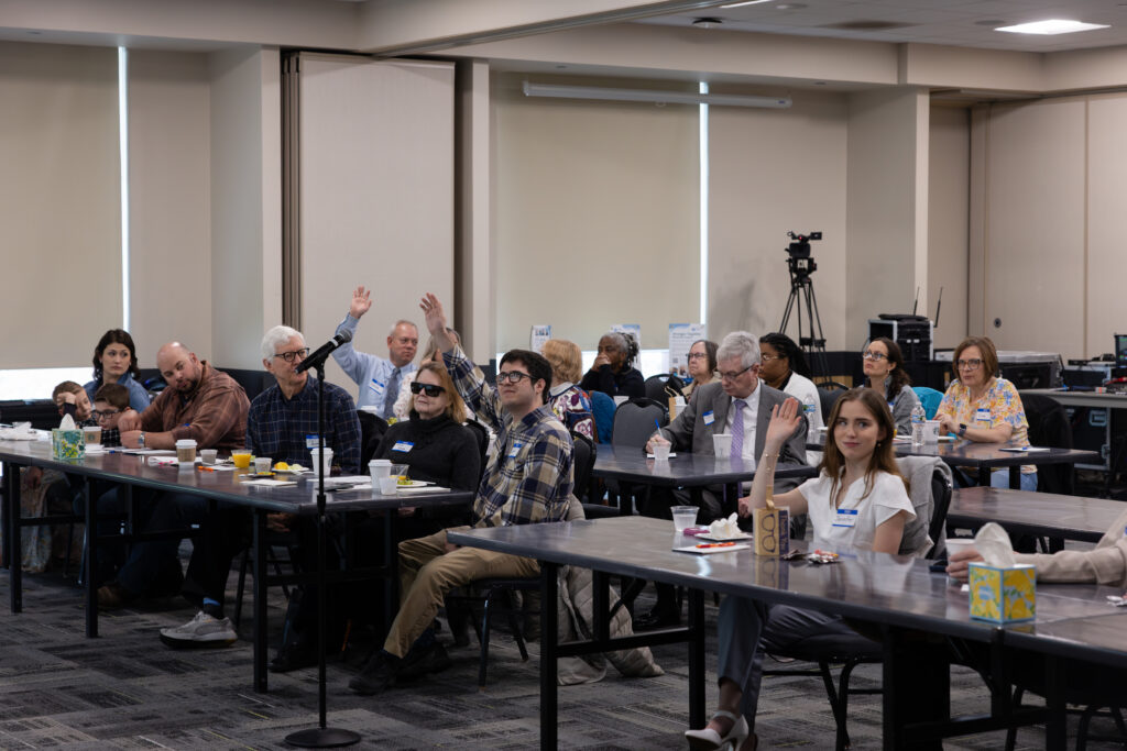
Families came ready with questions! Hands went up throughout the session as the conversation moved from FEVR to ROP, Best disease, Stickler syndrome, and beyond.
Adult ROP Is Real and Under-recognized
One audience member asked whether retinal issues continue to appear in adults who were treated for ROP as premature infants. The answer: yes, and this is a critically important topic as premature infant survival rates have dramatically improved.
Adults with a history of retinopathy of prematurity can experience:
- Retinal holes or tears
- Reactivation of abnormal blood vessels that leak fluid or lipid into the center of vision
- Slowly developing retinal detachments that can go unnoticed for years
Dr. Capone shared the story of a valedictorian who had spontaneously regressed ROP as an infant, never treated, and who came in as a young adult with a retinal detachment that had been progressing. He only noticed when it split his central vision.
💡 Critical Reminder for ROP Families: Patients with a history of ROP should be followed by a retina specialist for life. Detachments can develop slowly and without obvious symptoms, even in otherwise healthy, high-functioning young adults.
Laser vs. Injection: A Nuanced Answer
Dr. Trese addressed the shift in how ROP is treated in the NICU. Anti-VEGF injections (medications injected into the eye) have become more common in recent years because they’re easier to administer and less stressful for fragile newborns. But he was direct: laser remains the gold standard in his opinion.
Children treated with injections alone, without laser follow-up, can develop holes in peripheral areas of the retina that never developed proper blood vessels, and may need laser anyway down the road.
💡 For ROP Parents: If your child was treated with an injection for retinopathy of prematurity, ask your doctor whether follow-up laser or ongoing monitoring of the peripheral retina is part of the plan.
Patching: It’s Never Too Late At Any Age
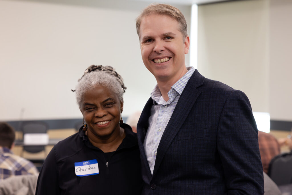
Charlene, a PRRF collaborator in the area of vision rehabilitation, poses with Dr. Trese.
One of the most memorable moments of the entire Q&A came from Dr. Patrick Droste, a decades-long collaborator of Dr. Michael Trese and former PRRF board member, who pushed back firmly on a common myth: that patching only works in young children.
“It is never too late to patch. Ever.” – Dr. Patrick Droste
Dr. Droste patches patients of all ages, including adults after retinal reattachment surgery, and consistently sees improved outcomes. His approach is practical and patient-centered:
- Start small. Even 20–30 minutes in the office to let the child and family get comfortable.
- Adjust to the child. The right amount of patching depends entirely on the individual.
- Trust the process. Every minute of patching reactivates retinal neurons and brain pathways that may have been dormant since the detachment.
Dr. Trese reinforced this: during his training, he would look at a severely distorted eye and assume the child could barely see, only to be told they were 20/100 or 20/200, in large part because parents had done the hard work of patching consistently.
🎧 Want to learn more? PRRF has a full podcast episode dedicated to patching. Visit this link to listen.
Best Disease Across Three Generations: Understanding Why the Same Gene Looks Different
An audience member asked one of the most emotionally resonant questions of the day: why does Best disease look so different across generations in the same family: a grandmother diagnosed in her 20s, a father with minimal vision loss, and a 5-year-old son already receiving injections?
“It seems so random,” she said. “Is it?”
Dr. Trese and Dr. Capone explained phenotypic variability: the same gene can produce dramatically different presentations of disease, even in the same family. The reasons aren’t fully understood yet, but likely involve complex interactions between multiple genes and a field called epigenetics, where genes can essentially be switched on or off.
💡 Research Opportunity: There is an active clinical trial for Best disease gene therapy at the Cincinnati Eye Institute, led by Dr. Robert Sisk. Families dealing with Best disease are encouraged to ask their physician about eligibility.
Stickler Syndrome: Surgical Advances While Genetic Research Grows
When it comes to Stickler syndrome, genetic therapy is not available yet. But researchers are actively working on it. The Pediatric Retinal Research Lab BioBank is being used to compile patient data and identify subtypes of Stickler and related vitreoretinal degenerations, many of which don’t yet have formal names in the medical literature.
In the meantime, surgical management has meaningfully advanced. Dr. Capone highlighted the use of methotrexate (an anti-scarring medication originally developed for cancer) during and after retinal detachment surgery in Stickler patients. Because children with Stickler face higher risk of a complication called proliferative vitreoretinopathy (PVR), where scar tissue forms inside the eye and causes repairs to fail, methotrexate has become an important tool for improving surgical outcomes over the last decade.
Clinical Trials for Pediatric Retinal Diseases: Should Your Family Consider One?
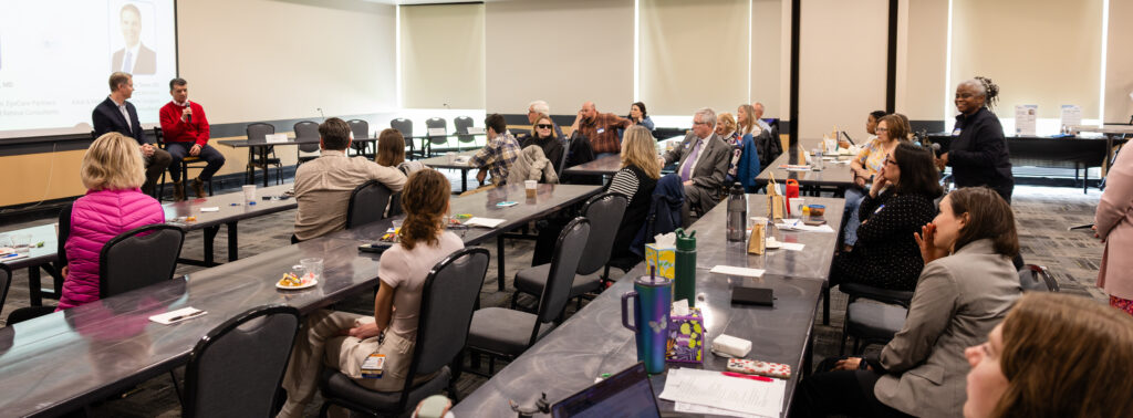
Dr. Capone gave a clear, accessible breakdown of how clinical trials work for families considering participation:
- Phase 1 — Safety: Small group, focused entirely on whether the treatment is safe.
- Phase 2 — Dosing: Larger group, testing a range of doses to identify effectiveness.
- Phase 3 — Confirmation: Large population, confirming safety and efficacy at the optimal dose.
- Phase 4 — Real World: Tracking outcomes after FDA approval, outside the trial setting.
His bottom line: clinical trials are designed with patient safety as the absolute top priority. All risks must be fully disclosed upfront. The phased structure exists specifically to catch problems before they reach a broad patient population.
⚠️ A Word of Caution: Dr. Capone noted that unregulated treatments given outside the clinical trial framework, such as stem cell injections into the eye, have caused serious, irreversible harm. The structure of a clinical trial is a protection, not red tape.
Gene Therapy for Pediatric Retinal Disease: The Horizon Is Getting Closer
Dr. Trese highlighted the remarkable pace of change. Luxturna (for RPE65 mutations in Leber congenital amaurosis (LCA)) has shown life-changing results and a new clinical trial for the most common form of LCA (CEP290) was recently announced.
The broader takeaway: the disease labels we’ve used for decades (FEVR, LCA, RP, Best disease) are increasingly being revealed as umbrella terms for many genetically distinct conditions. Personalized genetic medicine is the future of treatment for pediatric retinal diseases.
The Real Goal: Function, Independence, and a Full Life
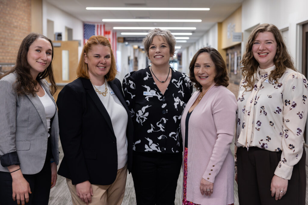
The PRRF behind the scenes team: working to ensure every family that contacts us feels seen, supported, and informed.
The session closed with a powerful reflection from an online audience member working in vision rehabilitation. She asked: “When do you know it’s time to shift from getting the patient to see a getting the patient to function?”
Dr. Trese’s answer: it’s never one or the other. The team always has function in mind, but timing matters. Introducing orientation and mobility training or cane use too early can feel to a patient like giving up. The goal is to keep the door open, raise the topic gently and consistently, and meet patients where they are.
“When you’re ready, we know the right people to send you to.” – Dr. Matt Trese
Watch the Full Session
This recap covers the highlights, but there’s so much more in the full conversation.
Watch the complete Q&A on YouTube →
—
The responses shared at this event are educational in nature and are not a substitute for individualized medical advice. Please consult your care team with questions specific to your child.
Visit PRRF.org to explore our podcast library, learn about our BioBank, and find resources for families living with pediatric retinal diseases.
Tags: FEVR, familial exudative vitreoretinopathy, FEVR retina, ROP, retinopathy of prematurity, adult ROP, Best disease, Stickler syndrome, LCA, Leber congenital amaurosis, gene therapy pediatric retina, retinal detachment children, pediatric retinal diseases, patching amblyopia, low vision children, smart glasses visually impaired, glasses for visually impaired, norrie disease, x-chromosomal retinoschisis, PRRF, Pediatric Retinal Research Foundation, clinical trials retinal disease, visually impaired children, glasses for blind people, vision impaired glasses








 Español de México
  English
  Français
  Deutsch
  Nederlands
  Português
  简体中文
简体中文  日本語
日本語  Русский
  Italiano