Familial Exudative Vitreoretinopathy (FEVR)
Introduction and History
Familial Exudative Vitreoretinopathy, or FEVR, is a rare genetic eye condition that affects the development of blood vessels in the retina, whereby the blood vessels at the edges of the retina don’t form properly. This can lead to a lack of blood supply and oxygen, causing the growth of new, abnormal blood vessels. These weak vessels can leak fluid (exudate) or bleed, sometimes leading to the formation of scar tissue, retinal detachment (where the retina pulls away from the back of the eye), and vision loss.
FEVR is “familial,” meaning it can be passed down through families. However, the severity of FEVR can vary significantly, even among family members. Some people may exhibit no symptoms, while others may experience severe vision problems.
FEVR was first identified and described as a distinct eye disease in 1969 by doctors Criswick and Schepens. They observed a pattern of abnormal retinal blood vessel development in several families, distinct from other known eye conditions.
Who Is Affected?
FEVR is a rare condition, and its exact frequency isn’t precisely known, partly because mild cases may go undiagnosed. It affects both males and females, though the pattern of inheritance can differ depending on the specific gene involved. FEVR can occur in people of all racial and ethnic backgrounds. Because it’s a genetic condition, individuals with a family history of FEVR are at a higher risk. The age at which FEVR is discovered can vary from early infancy to adulthood, though symptoms often begin in childhood.
Diagnosis
Diagnosing FEVR involves a careful eye examination by an ophthalmologist (eye doctor), often a specialist in retinal diseases. The doctor will look for characteristic signs in the retina, such as:
A key diagnostic test is wide-field fluorescein angiography, which highlights any abnormalities, such as missing vessels or leakage. Genetic testing can also help confirm the diagnosis by identifying mutations in genes known to cause FEVR, such as FZD4, LRP5, TSPAN12, and NDP. Examining family members can also be important.
Treatment
Treatment for FEVR aims to prevent the disease from worsening and to preserve vision. The specific treatment depends on how severe the condition is:
Prognosis
The outlook for individuals with FEVR varies greatly. With early diagnosis and timely treatment, vision can often be stabilized, and severe vision loss can be prevented in many cases. However, FEVR is a lifelong condition and can sometimes progress despite treatment, especially in more severe forms. Some individuals may experience significant vision impairment. Regular follow-up appointments with an eye doctor are crucial for monitoring the condition.
Current Research
Researchers are working diligently to gain a deeper understanding of FEVR and develop new treatments. Current research focuses on:
Continued research holds promise for even more effective ways to manage FEVR and preserve the vision of those affected.
References
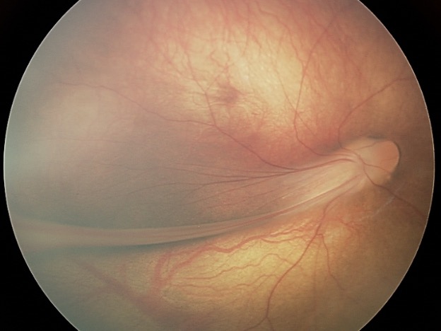
The retina is drawn into a fold by scar tissue.


© 2025 Pediatric Retinal Research Foundation.