Norrie Disease (ND)
Introduction and History
Norrie disease is a rare genetic disorder that primarily affects males, leading to blindness at birth and often hearing loss and developmental challenges as children grow. The condition was first described in 1961 by Danish ophthalmologist Mette Warburg, who observed a hereditary pattern of retinal degeneration in a family over seven generations. The condition is named after Gordon Norrie, another Danish ophthalmologist who observed similar cases in 1927. (JAMA Network, Wikipedia)
Who Is Affected?
Norrie disease is an X-linked genetic condition. This means the gene responsible for this disease is found on the X chromosome. Since males have only one X chromosome (they are XY) and females have two (they are XX), Norrie disease typically affects males more often. Females can carry the gene without showing symptoms or only have very mild symptoms because their other X chromosome functions normally.
Norrie disease is extremely rare. While the exact number of affected individuals is unknown, over 400 cases have been documented. It is estimated that the disorder occurs in approximately 1 in several hundred thousand people and can impact individuals from various ethnic backgrounds.
Diagnosis
Diagnosis is made when a baby shows signs of severe vision problems shortly after birth or clinical signs such as leukocoria (white pupil reflex), retinal detachment, and pseudogliomas (as in the figure). Genetic testing can confirm the presence of mutations in the NDP gene located on the X chromosome. Prenatal testing is possible if the mutation is known in the family. (AAO, Orpha.net). This test looks for the specific changes in the NDP gene. Genetic testing can identify the mutation in around 85% of affected males, helping doctors understand the condition and provide the best care [1].
Treatment
There is no cure for the profound vision loss caused by Norrie disease, but early intervention can improve quality of life. Surgical options like vitrectomy (removal of the vitreous gel) and lensectomy (removal of the lens) have shown benefits in preserving light perception and preventing further eye damage. For hearing loss, hearing aids and cochlear implants can significantly improve communication abilities. Supportive therapies, including special education and behavioral interventions, are crucial for addressing developmental and cognitive challenges. (JAMA Network, EBSCO)
Prognosis
With appropriate management, individuals with Norrie disease can lead fulfilling lives. However, the condition often leads to blindness and hearing loss, which can impact daily activities and development. Life expectancy is generally normal, but associated complications may require ongoing care.
Current Research
Recent studies are exploring gene therapy as a potential treatment for Norrie disease. In 2023, researchers demonstrated that neonatal gene therapy could prevent hearing loss and restore retinal function in animal models, offering hope for future treatments. (EBSCO)
References
- Warburg M. Hereditary retinal degeneration with congenital blindness. Acta Ophthalmol. 1961;39:1–25.
- Orphanet: Norrie disease. Available at: https://www.orpha.net/en/disease/detail/649
- MedlinePlus Genetics: Norrie disease. Available at: https://medlineplus.gov/genetics/condition/norrie-disease/
- Walsh MK, Drenser KA, Capone A Jr, Trese MT. Early vitrectomy effective for Norrie disease. Arch Ophthalmol. 2010;128(4):456-460.
- Todorich B, Thanos A, Yonekawa Y, Capone A Jr. Repair of Total Tractional Retinal Detachment in Norrie Disease. Ophthalmic Surg Lasers Imaging Retina. 2017.
- EBSCO Research Starters: Norrie disease. Available at: https://www.ebsco.com/research-starters/health-and-medicine/norrie-disease
- Pauzuolyte V, et al. Systemic gene therapy rescues retinal dysfunction and hearing loss in model of Norrie disease. EMBO Mol Med. 2023;15(10)
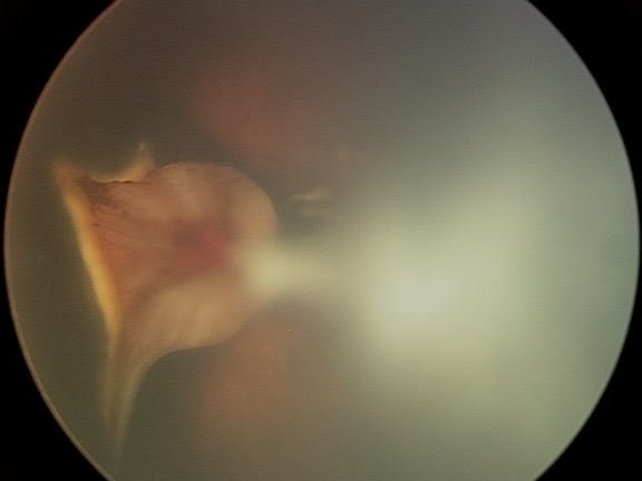
The poorly developed and disorganized retina appears as a whitish mass at the back of the eye. It is connected by a stalk to disorganized fibrovascular tissue at the front of the eye (out of focus).