Retinopathy of Prematurity (ROP)

Introduction and History

Retinopathy of prematurity (ROP), often called “ROP,” is an eye disease that can affect babies born too early (prematurely). It happens because the blood vessels in the retina haven’t finished growing when the baby is born. In ROP, these blood vessels grow abnormally and can lead to serious vision problems, including blindness, if left untreated.

The condition was first described in medical literature by Dr. Theodore Terry in 1942, who called it “retrolental fibroplasia” [1]. In the 1940s and 1950s, ROP caused an “epidemic” of blindness in premature babies, largely due to the unmonitored use of high levels of oxygen in incubators. Once doctors learned about the connection between oxygen and ROP, careful management of oxygen helped reduce cases. However, as medical care for premature babies improved and even very tiny infants began to survive, ROP unfortunately became a significant issue once again [2].

Who Is Affected?

ROP primarily affects premature infants born very early or with very low birth weights. The earlier a baby is born and the less they weigh at birth, the higher their risk of developing ROP.  Babies born before 30-31 weeks of pregnancy or weighing less than 3.3 pounds (1500 grams) are most at risk [3, 4]. While ROP affects infants of all racial and ethnic backgrounds, its incidence can vary globally. In some developing countries, where more premature babies are surviving, ROP is an increasing cause of childhood blindness [4].

Other factors that can increase the risk of ROP include breathing problems, infections, bleeding in the brain, and overall poor health after birth [4].

Diagnosis

ROP is diagnosed through a special eye exam conducted by an ophthalmologist (an eye doctor) who specializes in treating babies. These exams typically start a few weeks after birth for at-risk infants. During the exam, the doctor uses a special light and magnifying tool to examine the retina and check for abnormal blood vessel growth.

ROP is categorized into different “zones” based on the location of the abnormal vessels in the eye and “stages” (from 1 to 5) depending on the severity of the changes [2, 4]. A severe form, sometimes called “plus disease,” indicates that the condition is progressing rapidly [2]. Regular screening exams are crucial for catching ROP early, before it leads to permanent vision loss.

Treatment

Most cases of ROP (stages 1 and 2) are mild and often get better on their own without treatment. However, if ROP progresses to more severe stages (stage 3 and beyond), treatment is necessary to prevent retinal detachment and blindness.

  • Laser Therapy (Photocoagulation): This is a common treatment where a laser is used to make tiny burns on the outer, undeveloped edges of the retina. This stops the abnormal blood vessels from growing and often prevents further progression of the disease [4].
  • Anti-VEGF Injections: A newer treatment involves injecting a medication (called an anti-VEGF agent, like bevacizumab or aflibercept) directly into the eye. These medications work by blocking a protein that causes abnormal blood vessels to grow. Anti-VEGF injections can be particularly effective for more severe or aggressive forms of ROP [5, 6]. Aflibercept (Eylea) received FDA approval in February 2023 as the first pharmacologic treatment for ROP in preterm infants [7].
  • Surgery (Vitrectomy): If ROP is very severe and leads to a retinal detachment (Stage 4 or 5 ROP), surgery may be needed to reattach the retina. This complex surgery, called a vitrectomy, is performed by a retinal specialist [4]. Dr. Antonio Capone Jr. is a leading expert in pediatric retinal diseases and has contributed to the understanding and surgical management of severe ROP and similar conditions [8].

Prognosis

The good news is that with careful screening and timely treatment, many babies with ROP have good vision outcomes. However, even if ROP goes away or is successfully treated, premature babies who had ROP may still have a higher risk for other eye problems later in life, such as:

  • Myopia (nearsightedness)
  • Strabismus (crossed eyes)
  • Amblyopia (lazy eye)
  • Glaucoma (high pressure in the eye)
  • Late retinal detachments [2].

Regular follow-up eye exams are important throughout childhood and even into adulthood for those who had ROP.

Current Research

Research into ROP is actively ongoing to improve prevention, diagnosis, and treatment. Scientists are exploring:

  • Better Screening Methods: Researchers are looking into new ways, including telemedicine and artificial intelligence, to screen babies for ROP more effectively and reach more at-risk infants [9].
  • Understanding Risk Factors: Studies continue to identify specific risk factors, including genetic factors, that make some babies more likely to develop severe ROP [4].
  • New Drug Therapies: Scientists are studying different anti-VEGF medications and other potential drugs that could prevent or treat ROP with fewer side effects [6, 7].
  • Optimizing Oxygen Management: Research continues on the ideal levels of oxygen for premature infants to minimize the risk of ROP while ensuring healthy development.

These research efforts aim to reduce the impact of ROP and ensure the best possible vision for premature babies.

References
  1. Terry, T. L. (1942). Fibroblastic Overgrowth of Persistent Tunica Vasculosa Lentis in Infants Born Prematurely: II. Report of Cases-Clinical Aspects. Transactions of the American Ophthalmological Society, 40, 262–284.
  2. Bajaj, M., Sharma, V., & Singh, R. K. (2015). Retinopathy of prematurity: Past, present and future. World Journal of Ophthalmology, 5(4), 682–692.
  3. Orphanet. (n.d.). Retinopathy of prematurity. Retrieved from https://www.orpha.net/consor/cgi-bin/Disease_Search.php?lng=EN&data_id=649&search=Disease_Search_Simple&Chr=649&diseaseType=Pat&Disease(s)/group%20of%20diseases=Retinopathy-of-prematurity
  4. MedlinePlus. (2024, February 5). NDP gene. Retrieved from https://medlineplus.gov/genetics/gene/ndp/ (While this reference primarily discusses the NDP gene, it mentions its relevance to ROP and provides a good 8th-grade level overview of related conditions.)
  5. International Classification of Retinopathy of Prematurity, 3rd edition. (2024). Ophthalmology, 131(6), 661–677. (Antonio Capone, Jr. is an author on this publication.)
  6. Chang, Y. T., Weng, S. F., Lee, L. F., Su, C. Y., & Chen, J. Y. (2025). New Aspects on the Treatment of Retinopathy of Prematurity: Currently Available Therapies and Emerging Novel Therapeutics. International Journal of Molecular Sciences, 23(15), 8345.
  7. Medscape. (2023, February 9). Retinopathy of Prematurity Treatment & Management. Retrieved from https://emedicine.medscape.com/article/976220-treatment
  8. Trese, M. T., & Capone, A. Jr. (2014). Surgical management of retinopathy of prematurity. Current Opinion in Ophthalmology, 25(3), 195–200.
  9. Cherecheanu, A. P., Rata, M. N., Cherecheanu, M. E., & Dinescu, B. M. (2023). Latest Trends in Retinopathy of Prematurity: Research on Risk Factors, Diagnostic Methods and Therapies. Medicina, 59(3), 565.

Image of eye: The peripheral retina is not vascularized in this image, with stage 1 ROP (a “demarcation line” positioned between the vascularized and non-vascularized retina) and dilated, tortuous vessels characteristic of “plus disease”.

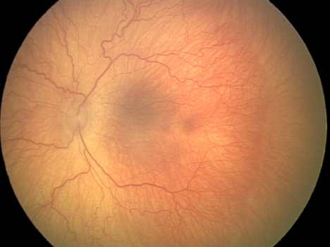
The peripheral retina is not vascularized in this image, with stage 1 ROP (a “demarcation line” positioned between the vascularized and non-vascularized retina) and dilated, tortuous vessels characteristic of “plus disease”.

en_USEnglish